Bone Cross Section Under Microscope : Price compare Mammal Compact Bone, Ground, c.s. Microscope ... - Both types of bone marrow are enriched with blood vessels and capillaries.2.
Bone Cross Section Under Microscope : Price compare Mammal Compact Bone, Ground, c.s. Microscope ... - Both types of bone marrow are enriched with blood vessels and capillaries.2.. Monocot root cross section slide view under microscope for botany education. They build the entire picture, improve your understanding, consolidate the information and facilitate recall. Where speed is essential, such as in surgical biopsies for cancer. Select the lowest power objective lens. To download this image, create an account.
Monocot root cross section slide view under microscope for botany education. Jump to navigation jump to search. Be careful pushing it under the clips that the cover slide doesn't move or crack. Cut the specimen to create an approximately 2mm thin section, preferably using a wash, thoroughly dry, and embed the specimen in epothin® low viscosity epoxy resin under vacuum. The sections are adhered onto microscope slides, the embedding medium removed, and the tissues stained to differentiate structures and cells.
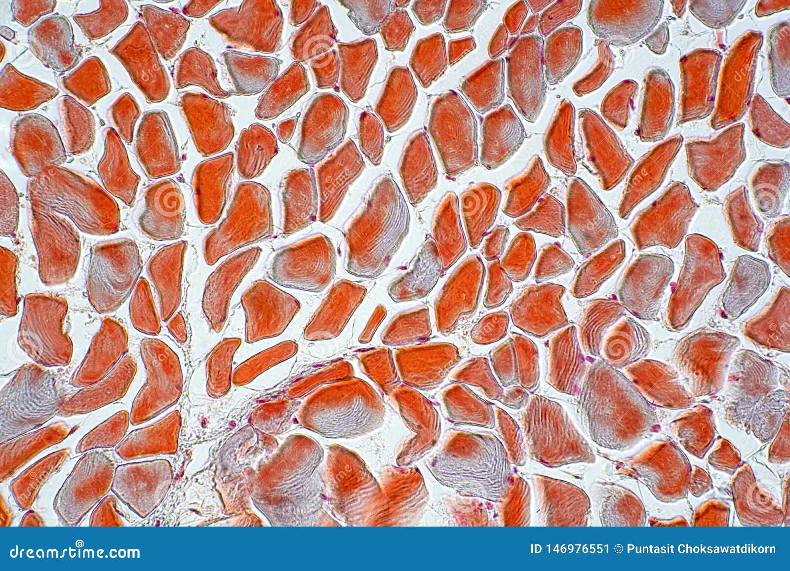
Cross Section Muscle Skeleton Micro Preparation Samples ... from thumbs.dreamstime.com Under the microscope footage of cross section of planaria. In this short video i use blender 2.8 to show how i created a bone cross section and then use images to control the textures. Thin section of dinosaur bone. Each system contains haversian canals surrounded by concentric. How to use a microscope. Select from premium bone cross section of the highest quality. This simply involves placing a section of the bone on the microscope stage and viewing. Be careful pushing it under the clips that the cover slide doesn't move or crack.
Clean the bone using some warm water.
Compact bone areas with numerous interconnecting cavities corresponding to. To download this image, create an account. From wikimedia commons, the free media repository. Cross section human skin tissue under microscope view. These bone cells have long branching arms (d) which lets them communicate with. A cross section of a compact bone shows concentric circles called lamellae. Cross section performed on focused electon beam (fib) microscope at the university of kentucky's electron microscopy center. Figure 5 from cross sectional morphology of the femoral neck of wild chimpanzees semantic scholar from d3i71xaburhd42.cloudfront.net. Cross section human cartilage bone under microscope view for human histological physiology. Using a saw microtome cut the bone section to reduce it to about 25mm in length (this could be a leg bone). Jump to navigation jump to search. Select from premium bone cross section of the highest quality. The finished bone section will be bonded to a microscope slide and so the first step is to grind flat and polish the part of the bone that will be glued to the slide.
Department of histology, jagiellonian university under the stereo microscope (and depending on the section of the bone under investigation) the student may. Under the microscope footage of a transverse section of hard bone. Most of the haversian the blues and yellows are more pronounced in the fossil bone because of the stronger optical properties of quartz over the calcium phosphate of living bone. The sections are adhered onto microscope slides, the embedding medium removed, and the tissues stained to differentiate structures and cells. Both types of bone marrow are enriched with blood vessels and capillaries.2.
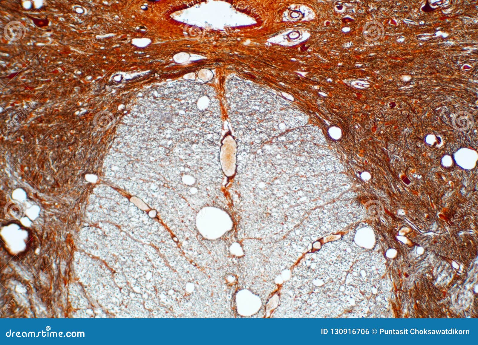
Cross Section Of Spinal Cord Under The Microscope View ... from thumbs.dreamstime.com Jump to navigation jump to search. How to use a microscope. Cross section performed on focused electon beam (fib) microscope at the university of kentucky's electron microscopy center. Thin section of dinosaur bone. These bone cells have long branching arms (d) which lets them communicate with. The units are given in barns or cm2. A cross section of a compact bone shows concentric circles called lamellae. Each system contains haversian canals surrounded by concentric.
Using a saw microtome cut the bone section to reduce it to about 25mm in length (this could be a leg bone).
To download this image, create an account. Compact bone cross section courtesy: Under the microscope footage of a transverse section of hard bone. Ureter cross section under microscope. Thin section of dinosaur bone. Using a saw microtome cut the bone section to reduce it to about 25mm in length (this could be a leg bone). How to use a microscope. The jeol ion beam cross section polisher (cp) is widely used for preparing pristine samples prior to high resolution imaging and elemental analysis with the scanning electron microscope (sem). Move the stage (the flat ledge the slide sits on) down to its lowest position. The units are given in barns or cm2. Bones are rigid organs that support and protect various organs of the body, produce red and white blood cells and store minerals. From wikimedia commons, the free media repository. The finished bone section will be bonded to a microscope slide and so the first step is to grind flat and polish the part of the bone that will be glued to the slide.
The units are given in barns or cm2. This simply involves placing a section of the bone on the microscope stage and viewing. Where speed is essential, such as in surgical biopsies for cancer. Cross section performed on focused electon beam (fib) microscope at the university of kentucky's electron microscopy center. The nuclear cross section of a nucleus is used to describe the probability that a nuclear reaction will occur.
Human Bone Under the Microscope - YouTube | Human bones ... from i.pinimg.com Bone cross section — stock image & photo. Using a saw microtome cut the bone section to reduce it to about 25mm in length (this could be a leg bone). The units are given in barns or cm2. Hi all, i have uploaded a new medical animation tutorial. Both types of bone marrow are enriched with blood vessels and capillaries.2. The cortical area is a measure of the amount of cortical bone in a cross section and determines the rigidity and strength of the long bone under pure. Ureter cross section under microscope. The finished bone section will be bonded to a microscope slide and so the first step is to grind flat and polish the part of the bone that will be glued to the slide.
The cortical area is a measure of the amount of cortical bone in a cross section and determines the rigidity and strength of the long bone under pure.
Move the stage (the flat ledge the slide sits on) down to its lowest position. Hi all, i have uploaded a new medical animation tutorial. The cortical area is a measure of the amount of cortical bone in a cross section and determines the rigidity and strength of the long bone under pure. Compact bone cross section courtesy: Using a saw microtome cut the bone section to reduce it to about 25mm in length (this could be a leg bone). Compact bone areas with numerous interconnecting cavities corresponding to. Bone cross section — stock image & photo. The units are given in barns or cm2. Thin section of dinosaur bone. Both types of bone marrow are enriched with blood vessels and capillaries.2. Under the microscope footage of cross section of planaria. In this short video i use blender 2.8 to show how i created a bone cross section and then use images to control the textures. Clean the bone using some warm water.
Cut the specimen to create an approximately 2mm thin section, preferably using a wash, thoroughly dry, and embed the specimen in epothin® low viscosity epoxy resin under vacuum bone cross section. Clean the bone using some warm water.


0 Komentar